 Lävi
Lävi Clinical Suite · Longitudinal brain MRI
Not every difference is disease.
Lävi Delta is in development and is not CE-marked.
We develop technology for detecting and assessing meaningful change between serial brain MRI examinations. Lävi Delta, our first product, supports the radiologist in follow-up brain MRI: it shows on a map where the signal has changed between two examinations. When the two cannot be compared on technical grounds, it says so and withholds the result.
Clinical validation and regulatory development in progress · not CE-marked
Baseline

Follow-up

Follow-up + change map

Selected slices
Public research data (MSLesSeg; Yale-Brain-Mets-Longitudinal, TCIA). CC BY 4.0. Skull removed, change map added.
Data and licences
Data: Guarnera, F., Rondinella, A., Crispino, E., Russo, G., Di Lorenzo, C., Maimone, D., Pappalardo, F., & Battiato, S. (2025). MSLesSeg: baseline and benchmarking of a new Multiple Sclerosis Lesion Segmentation dataset [Dataset]. figshare. https://doi.org/10.6084/m9.figshare.27919209.v1 · Licence: CC BY 4.0.
Data: Chadha, S., Weiss, D., Janas, A., Ramakrishnan, D., Hager, T., Osenberg, K., Willms, K., Zhu, J., Chiang, V., Bakas, S., Maleki, N., Sritharan, D. V., Schoenherr, S., Westerhoff, M., Zawalich, M., Davis, M., Malhotra, A., Bousabarah, K., Deuschl, C., Lin, M., Aneja, S., & Aboian, M. (2025). Yale longitudinal dataset of brain metastases on MRI with associated clinical data (Yale-Brain-Mets-Longitudinal) (Version 1) [Dataset]. The Cancer Imaging Archive. https://doi.org/10.7937/3YAT-E768 · Licence: CC BY 4.0.
Clark, K., Vendt, B., Smith, K., Freymann, J., Kirby, J., Koppel, P., Moore, S., Phillips, S., Maffitt, D., Pringle, M., Tarbox, L., & Prior, F. (2013). The Cancer Imaging Archive (TCIA): maintaining and operating a public information repository. Journal of Digital Imaging, 26(6), 1045–1057. https://doi.org/10.1007/s10278-013-9622-7
Images have been processed: skull removed, change map added.
Public research data, not patient data from our study. Lävi Delta is in development and is not CE-marked.
01 — What it does
What Lävi Delta does
Lävi Delta compares a person's new brain MRI with their previous one and shows, on a colour map, where the signal has changed. The radiologist checks each coloured area against the original images and decides what it means; the software makes no diagnosis. When two examinations cannot fairly be compared, it says so and shows no result rather than a misleading one.
- Red: signal increased between the two scans.
- Blue: signal decreased.
- No colour: the map shows no change. The stronger the colour, the larger the change.
The map is not a list of lesions and not a diagnosis. The radiologist decides.
02 — Scanners
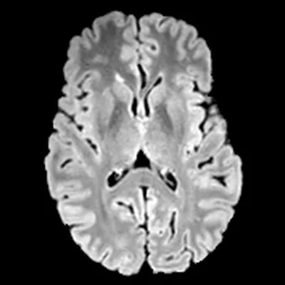
Same person, two scanners
A follow-up scan is often made on a different scanner from the first one, and some of what looks like change is only the difference between the two machines. Lävi Delta is being built to tell that apart from real change in the patient.
Scanner 1 · Philips
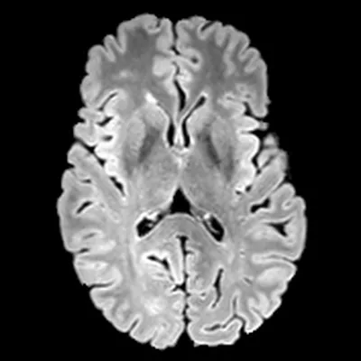
Scanner 2 · Siemens
Plain subtraction
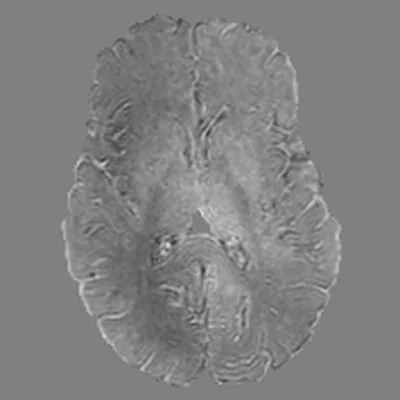
Plain subtraction: grey means the two scans agree; lighter or darker means they differ.
Red: signal increased between the two scans. Blue: signal decreased. The stronger the colour, the larger the change. Where there is no colour, the map shows no change.
Yellow rings: simulated findings.
In our internal technical tests, results across scanners from different vendors have been promising. Read what was tested and what was not.
03 — Development status
Where we are
Fig. 3 — Where we arestages, not dates
Lävi Delta is in development and is not CE-marked.
What exists today, and what is not yet established → The planned reader study →
Status as of 2026-11-17
04 — Contact
Talk to us
One address for everyone — the subject line tells us who is writing. Andreas, the CEO, answers his own email.
-
Hospitals
If your department reads follow-up brain MRI routinely, we can talk through what a pilot would involve. Today a pilot is a research collaboration inside your own network, not clinical use. What a pilot involves.
-
Partners and investors
The evidence plan and the technical architecture, examined in detail. We maintain dated internal development, validation and evidence-planning documentation; selected material can be shared in qualified conversations under NDA or controlled data-room access.
Research or technology partner? Write about research collaboration.
-
Radiologists
What the change map shows, how it would fit a follow-up read, and what it does not do. A neuroradiologist answers.
Looking for work, or interested in MS imaging? Careers · Lävi MS · The company and team